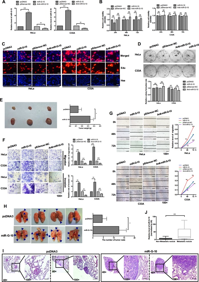
Fig. 2. miR-G-10 promotes oncogenic activity in cervical cancer cells.
a Relative level of miR-G-10 after transfection with miR-G-10 or Anti-miR-G-10 in HeLa and C33A. b MTT assay, c EdU assay (scale bar: 20 μm), and d colony-formation assay (scale bar: 5 mm) were used to detected cell proliferation. e Representative graph of tumors (left) and tumor weights (right). f Transwell assays showed the invasion tendency. Scale bar: 50 μm. g Wound-healing assays were used to measure migration. Scale bar: 50 μm. h In vivo metastasis assay. Representative lungs were harvest after injecting HeLa cells with pcDNA3 or miR-G-10 overexpression. i Representative H&E staining results of metastatic nodules in the lungs are shown. Scale bar: 50 μm. j miR-G-10 levels in serum from cervical cancer patients with/without metastasis. The expression levels were normalized to U6 snRNA. All of the experiments were repeated three times. *P < 0.05; **P < 0.01; ***P < 0.001. NS not significant

