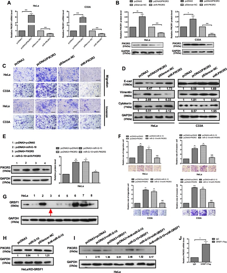
Fig. 5. GRSF1-mediated miR-G-10 upregulates the PIK3R3 and promotes the malignant phenotype of cervical cancer cells.
a, b RT-qPCR and western blotting showed PIK3R3 levels in HeLa C33A cells with overexpress or knockdown plasmid. c Transwell assays showed that PIK3R3 promotes cells’ migration and invasion. Scale bar: 50 μm. d Western blotting showed the protein levels of E-cadherin, vimentin, and cytokeratin after transfection with PIK3R3 or shR-PIK3R3. e Co-transfection with miR-G-10 and PIK3R3 showed that PIK3R3 partly rescued the upregulation of miR-G-10 on PIK3R3 and f cell migration and invasion in HeLa and C33A. Scale bar: 50 μm. g Western blot assay was used to determine the protein level of GRSF1-knockdown cells (#1-#8) and HeLa cell as a control. h We examined the protein level of PIK3R3 in GRSF1-knockdown cells (#3) by western blot after transfection miR-G-10 and anti-miR-G-10. i Western blot assay showed the expression levels of PIK3R3 transfected with the indicated plasmids in GRSF1-knockdown cells. j RT-qPCR showed the RNA level of PIK3R3 in the GRSF1-RIP complex. All of the experiments were repeated three times. *P < 0.05; **P < 0.01; ***P < 0.001. NS not significant

