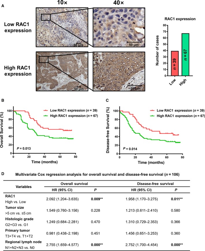
Figure 1.

IHC staining of RAC1 and prognostic significance evaluation in ESCC patient samples. (A) IHC detection of RAC1 expression in 106 ESCC patient samples. (B, C) Kaplan–Meier survival analysis with log‐rank test evaluating the OS and DFS in patients expressing high or low levels of RAC1. (D) Multivariate Cox regression models for OS and DFS were performed. All scale bars, 50 μm.
