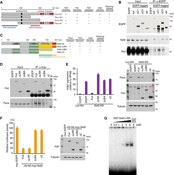
Schematic of full‐length Panx protein and deletion mutants. Predicted NLS and α‐helices and β‐strands are indicated (left panel). The results of rescue experiments are summarized (right panel).
Western blot (WB) of EGFP‐Immunoprecipitation (IP) product from lysate of EGFP‐tagged Panx deletion construct‐expressing OSCs, using EGFP, Nxf2, and Piwi antibody.
Schematic of full‐length Nxf2 and deletion mutant proteins. LRR: leucine‐rich repeat domain, NTF2‐like: nuclear transport factor 2‐like domain, UBA: ubiquitin‐associated domain (left panel). The results of rescue experiments and tethering assay are summarized (right panel).
IP using anti‐myc antibody from lysate of OSCs expressing myc‐tagged Nxf2 proteins, followed by WB using anti‐myc or anti‐Panx antibody.
mdg1 expression levels were monitored by qRT–PCR in OSCs expressing exogenous Nxf2 proteins treated with either siLuc (control) or siNxf2 (left panel). Error bars indicate SD (n = 3). Exogenous Nxf2 mRNAs are resistant to siRNA for Nxf2. Expression levels of Panx‐ and myc‐tagged Nxf2 proteins were confirmed by WB using anti‐myc and anti‐Panx antibodies. Tubulin was used as a loading control. Asterisk indicates the background signal (right panel).
Effects of λN fusion Nxf2 proteins on luciferase activity at 48 hpt. Error bars indicate SD (n = 3) (left panel). Expression levels of exogenous λN‐HA–myc‐Nxf2 proteins (right panel).
Electrophoretic mobility shift assay (EMSA) showing the binding of the first LRR region of Nxf2 (1–285 aa) to single‐stranded RNA. The indicated amount of recombinant protein was mixed with 1 nM single‐stranded RNA (16 nt).

