
Figure 4.
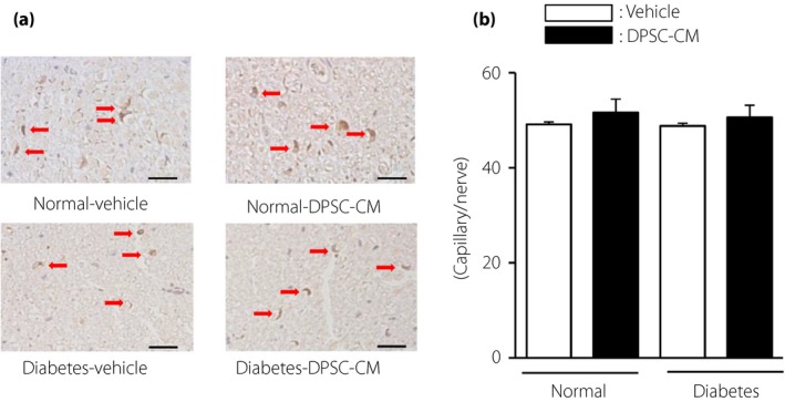
The number of capillaries in sciatic nerves was unaffected by dental pulp stem cell‐conditioned media (DPSC‐CM). (a) Representative photomicrographs of immunohistological staining of the sciatic nerves of normal and diabetic rats. Capillaries were visualized with platelet endothelial cell adhesion molecule 1. Scale bar, 10 μm. (b) Quantitative analysis of the number of capillaries in sciatic nerves of normal and diabetic rats (n = 4). The results are presented as the mean ± standard error of the mean.