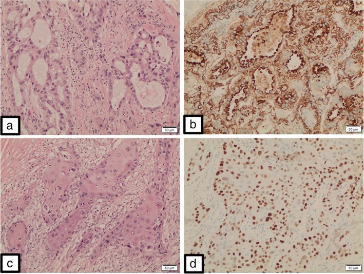
Figure 2.

Malignant glandular component made up of dysplastic cells arranged in distinct confluent glandular‐cribriform clusters (a). These neoplastic glandular elements are positive for Napsin A (b) and negative for p63. Malignant squamous component made up of polygonal tumor cells arranged in solid infiltrative clusters (c). Individual cell keratinization and intercellular bridges are clearly evident. These neoplastic squamoid elements are positive for p63 (d) and negative for Napsin A. (x100).
