
Figure 5.
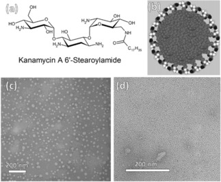
(a) Molecular structure of Kanamycin A 6′‐Stearoylamide, (b) a model of the micelle made of Kanamycin A 6′‐Stearoylamide in solution, and transmission electron micrographs (TEMs) of (c) micelles deposited from a dilute Kanamycin A 6′‐Stearoylamide (10‐5 M) and (c) a 100‐fold concentration on a carbon grid. The diameter of the micelles in (d) is a uniform 6 nm, corresponding to a fluid bilayer of Kanamycin A 6′‐Stearoylamide. Reprinted from21 with permission of American Chemical Society.