Fig. 1.
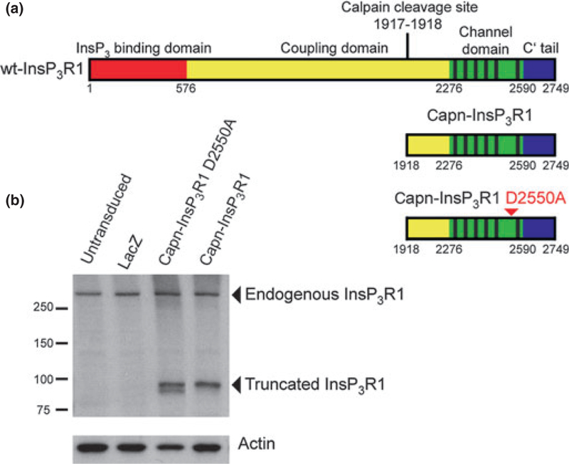
Schematic representation and expression of recombinant InsP3R1 constructs in primary neurons. (a) Protein domain structure of wt-InsP3R1 (top), calpain-cleaved InsP3R1 recombinant (middle), and calpain-cleaved InsP3R1 with the D2550A point mutation (bottom; arrowhead). Residues numbered according to rat type 1 SI+, SII+, SIII-sequence (protein accession NP_001007236.1). (b) Western blot analysis of whole-cell lysates from untransduced rat primary cortical cultures and primary cortical cultures transduced with AAV 2/1 expressing lacZ, capn-InsP3R1 D2550A, or capn-InsP3R1 at 1 week post-transduction (14 DIV). Carboxyl-terminal InsP3R1 antibody was used to detect endogenous and recombinant truncated rat InsP3R1. Antibody against actin was used as a loading control.

