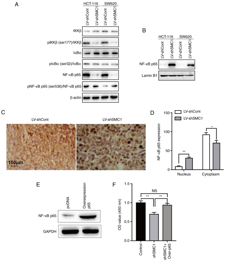
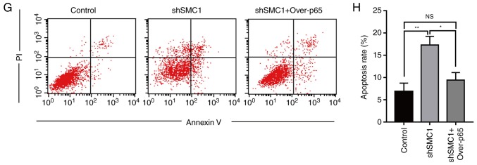
Figure 5.
Effects of SMC1 expression on the activity of the NF-κB signaling pathway in colorectal cancer cells. (A) Protein expression was examined in cytoplasmic cells lysates via western blotting. (B) Protein expression was examined in nuclear cells lysates by western blotting. (C and D) NF-κB p65 expression in xenograft tissue was determined via immunohistochemistry analysis. (E) SW620 cells were transfected with p65-overexpression plasmids, and NF-κB p65 protein levels were detected via western blotting. (F) Viability of SW620 cells at 96 h after knockdown of SMC1 and overexpression of p65. Cell viability was determined using an MTT assay. (G and H) Apoptosis was determined via flow cytometry in SW620 cells at 48 h following SMC1 knockdown and p65 overexpression. *P<0.05, **P<0.01. Cont, control; IκBα, inhibitor of nuclear factor-κB subunit α; IKKβ, inhibitor of nuclear factor-κB subunit β; LV, lentivirus; NC, negative control; NS, not significant; OD, optical density; Over-p65, overexpression of p65; p, phosphorylated; PI, propidium iodide; sh, short hairpin (RNA); SMC1, structural maintenance of chromosomes 1.


