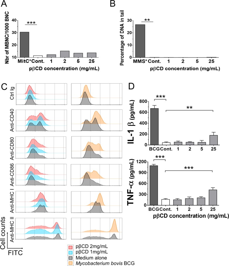
Figure 3.
pβCD are not genotoxic and poorly induce pro-inflammatory responses. (A, B) THP1 cells were incubated for 24 h with different concentrations of pβCD for the evaluation of genotoxicity. The micronucleus assay (A) was used to detect any damage that occurred during cell division (mitomycin was used as positive control), while the comet assay (B) was used to evaluate DNA strand breaks (methyl methanesulfonate was used as positive control). (C) BMDC were incubated overnight with pβCD or were inoculated with BCG (MOI = 1), as a positive control. After overnight incubation, cells were stained with FITC-conjugated antibodies. The FITC signal gated on CD11c+ cells shows the surface expression level of co-stimulatory molecules (CD40, CD80, CD86) and MHC-I or MHC-II molecules, as phenotypic dendritic cell maturation markers. (D) As a hallmark of functional dendritic cell maturation, IL-1β and TNF-α were quantified by ELISA in the supernatants of the same cultures, described in (C). Symbols ** and *** denote p < 0.01 and p < 0.001, respectively.

