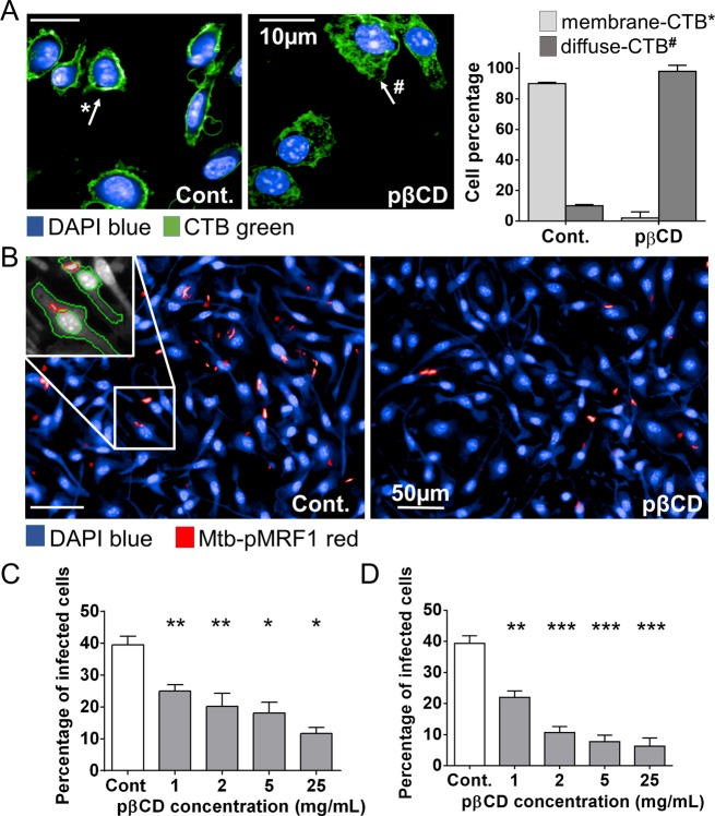
Figure 4.
pβCD disturb lipid raft integrity of macrophages and prevent Mtb uptake. BMDM were incubated overnight with pβCD at different concentrations and then fixed with formalin. (A) Effect of pβCD on cholesterol distribution at the plasma membrane in BMDM. Typical images obtained by automated confocal microscopy (pβCD: 16 h at 2 mg/mL) and related quantification. CTB-FITC- and DAPI-labeled nuclei shown in green and blue, respectively. Scale bar: 10 μm. (B, C, D) BMDM were incubated with different concentrations of pβCD for 2 or 16 h prior to infection with red-fluorescent Mtb. (B) Typical images of H37Rv-pMRF1- and DAPI-labeled nuclei are shown in red and blue, respectively (pβCD: 16 h at 5 mg/mL). Scale bar: 50 μm. Percentage of Mtb-infected BMDM (2 h (C) or 16 h (D) postinfection) upon pre-incubation with pβCD. Data are presented as mean ± SEM and are representative of two independent experiments. Symbols *, **, and *** denote p < 0.05, p < 0.01, and p < 0.001, respectively.

