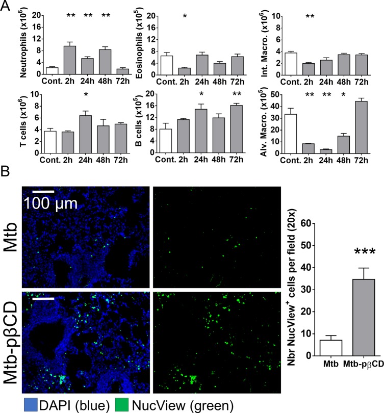
Figure 6.
pβCD induce alveolar macrophage depletion and pulmonary apoptosis. (A) BALB/c mice received one i.n. administration of pβCD (150 mg/mL, 50 μL) and were euthanized at indicated time points postadministration to evaluate the number of different cell populations in lungs by flow cytometry. The following cell types were analyzed: alveolar macrophages (CD11c+ F4/80+ SiglecF+), interstitial macrophages (F4/80+ CD11cint SiglecF–), neutrophils (CD11b+ LY6G+), eosinophils (SiglecF+ CD11c–), T cells (CD3+), and B cells (B220+ MHCII+). (B) BALB/c mice were inoculated via the i.n. route with Mtb H37Rv (105 CFU). At days 7, 9, 11, 14, 16, and 18 postchallenge, mice received administrations of 50 μL of pβCD (150 mg/mL) via the e.t. route. At day 21 postchallenge, 50 μL of NucView 488 caspase-3 substrate (diluted in PBS) was administered i.n. to each mouse for 1 h before lung harvesting and subsequent fluorescence histology analysis. Data are presented as mean ± SEM and are representative of two independent experiments. Symbols *, **, and *** denote p < 0.05, p < 0.01, and p < 0.001, respectively.

