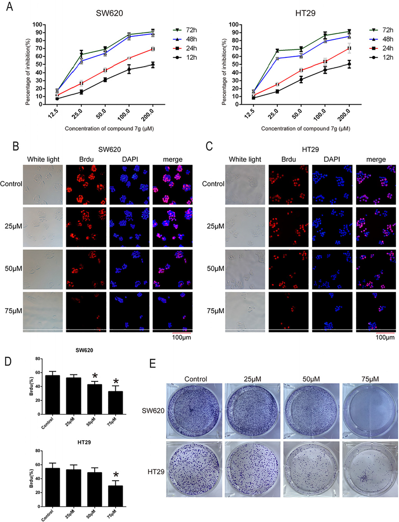
Fig. 1.
Compound 7g decreased proliferation and viability of colorectal cancer cell lines SW620 and HT29. (A) Cell proliferation and GI50 were assessed by MTT. SW620 and HT29 cells were treated with compound 7g at different concentrations (0, 12.5, 25, 50, 100, 200 μM) for 12 h, 24 h, 48 h, 72 h respectively, and then cell viability and GI50 were measured and calculated. Data are the mean ± SD of three independent experiments, and each experiment was conducted in sextuplicate. (B, C) BrdU immunofluorescence staining was conducted in SW620 and HT29 cell lines after treatment with compound 7g. The nuclei of SW620 and HT29 were exposed to compound 7g at 0, 25, 50, and 75 μM for 48 h, and then stained with BrdU (red fluorescence), primary mouse antibody against BrdU and anti-mouse IgG secondary antibody. Fluorescence microscopy was conducted after incubating with DAPI (blue fluorescence). The bar length for the group as shown in the panels is 100 μm. (D) The percentage of positive BrdU from panel B and C were analyzed, respectively. Every value was the average of three independent experiments. Data were shown as the mean ± SD. * = p < 0.05; ** = p < 0.01. (E) The effects of compound 7g on the clonogenicity of SW620 and HT29 were analyzed using the colony formation assay.

