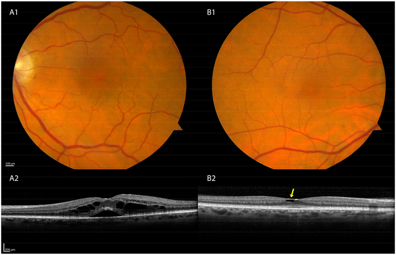
Figure 2: Less common disorders seen in OCT screening of normal eyes.
A1–2. Intraretinal fluid and subretinal fluid most likely represents post-operative cystoid macular edema (Irvine-Gass). B1–2. Eye with Macular Telangiectasia Type 2 has architectural cavitation with draping of the overlying internal limiting membrane. Fundus photographs are unremarkable.

