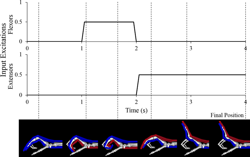
Figure 1:
Excitation inputs and resulting index finger motion for one of the four second forward dynamic simulations completed in this study (nominal model, 50% excitation level). Uniform excitation inputs were defined for the two extrinsic flexors of the index finger and the two extrinsic extensors. In these images, muscle-tendon paths that are blue indicate the muscles are passive, or “off” (0% excitation); paths that are red indicate the muscles are active, or “on” (50% excitation in this example). Dashed lines indicate the time at which the index finger reached the pictured position during the simulation, final dashed line indicates the final position used for the determination of the claw finger deformity.

