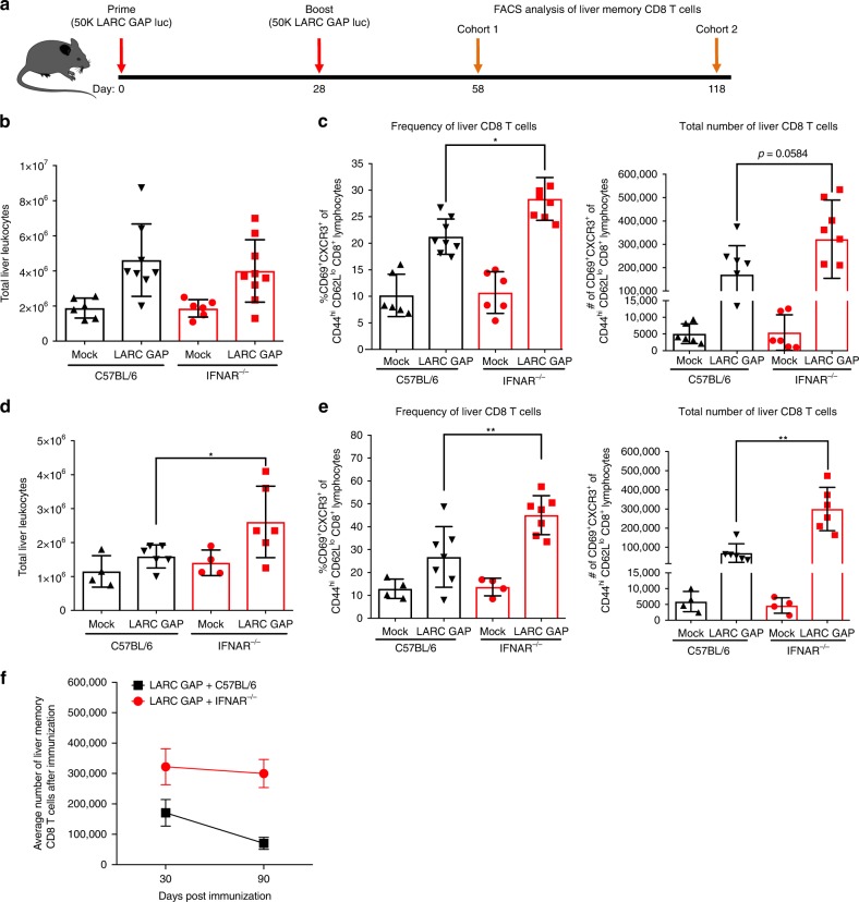
Fig. 3.
Lack of type I IFN signaling enhances the generation of liver memory CD8 T cells in LARC GAP-immunized mice. a Schematic of the immunization regimen. B6 mice and IFNAR−/− mice were immunized twice with 50,000 LARC GAP sporozoites. Thirty or 90 days after the second immunization liver non-parenchymal cells were isolated and subjected to FACS analysis. b Quantification of liver leukocytes isolated from immunized B6 mice and immunized IFNAR−/− mice, 30 days after the last immunization. c Analysis of liver memory CD8 T cells 30 days after GAP immunization. Liver memory CD8 T cells are defined as CD44hiCXCR3+CD69+ of CD62Llo CD8+CD4−CD3+ T lymphocytes. d Quantification of liver leukocytes isolated from immunized B6 mice and immunized IFNAR−/− mice, 90 days after the last immunization. e Analysis of liver memory CD8 T cells 90 days after immunization. Liver memory CD8 T cells are defined as CD44hiCXCR3+CD69+ of CD62Llo CD8+CD4−CD3+ T lymphocytes. f Comparison of the mean liver memory CD8 T cell numbers 30 or 90 days after boost. Data from panels b through f are compiled from at least two independent experiments with two mock immunized and at least three LARC GAP immunized mice in each group. Each dot represents a single mouse. Bar graphs are expressed as mean ± SD. *p < 0.05 and **p < 0.005 (from unpaired two-tailed Student’s t-test). Components of this figure were created using Servier Medical Art templates, which are licensed under a Creative Commons Attribution 3.0 Unported License; https://smart.servier.com

