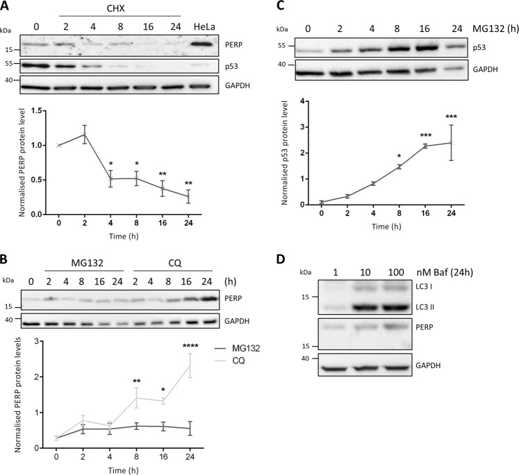
Fig. 4. PERP protein is actively degraded by the lysosome.
a HCT116 cells were treated with 30 μg/ml CHX for the indicated time points, and the protein levels of PERP and p53 were detected by immunoblotting. Histogram shows PERP protein levels normalized to GAPDH. One-way ANOVA, n = 3, F = 12.63, p = 0.0003***. b HCT116 cells were treated with 20 μM MG132 or 100 μM CQ for the indicated time points and PERP protein levels were detected by Western blot. Graph represents PERP protein levels normalized to GAPDH. One-way ANOVA, n = 3, MG132 PERP: F = 0.8849, p = 0.5204 ns; CQ PERP: F = 13.03, p = 0.0002***. c HCT116 cells were treated with 20 μM MG132 and the protein levels of p53 were detected by immunoblotting and normalized to the level of GAPDH. One-way ANOVA, n = 3, F = 11.58, p = 0.0003***. d HCT116 cells were treated with 1, 10 and 100 nM of Baf and the protein levels of LC3B and PERP were detected 24 h post-treatment by immunoblotting

