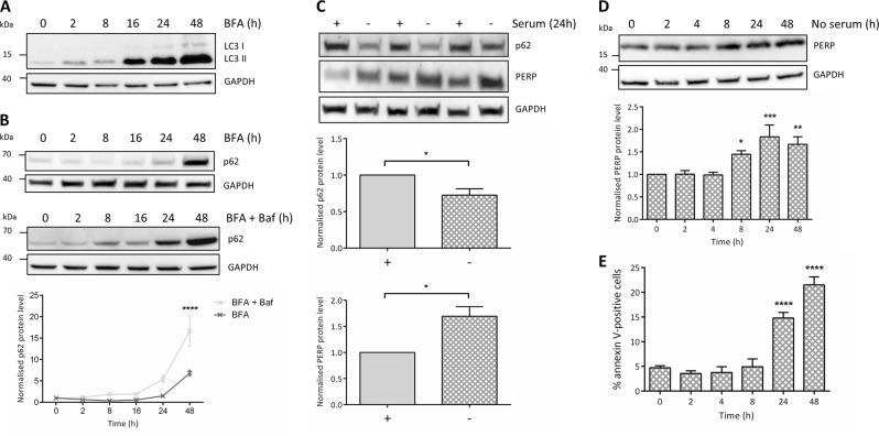
Fig. 5. PERP is selectively upregulated in response to sustained autophagy induction, correlating with apoptosis.
a Protein levels of autophagy marker LC3B in HCT116 cells treated with 1 μg/ml BFA. b Autophagy flux analysis of p62 levels in HCT116 cells treated with 1 µg/ml BFA in the presence and absence of 10 mM Baf. Protein levels quantified by densitometry and normalized to the level of GAPDH. Two-way ANOVA with Bonferroni post hoc test, n = 3. c HCT116 cells were cultured in the presence (+) or absence (−) of serum for 24 h and p62 and PERP protein levels were detected and normalized to the level of GAPDH. Blot from one independent experiment shown, performed in technical triplicate. Student’s t-test, n = 3, p62: p = 0.0361*, PERP: p = 0.02*. d HCT116 cells were serum starved for the indicated time points and PERP protein levels were detected by Western blot and normalized to the level of GAPDH. One-way ANOVA, n = 5, F = 7.426, p = 0.0003***. e HCT116 cells were serum-starved for the indicated time points and apoptosis induction was measured by flow cytometry using an Alexa Fluor 647 annexin V conjugate. One-way ANOVA, n = 4, F = 41.00, p < 0.0001****

