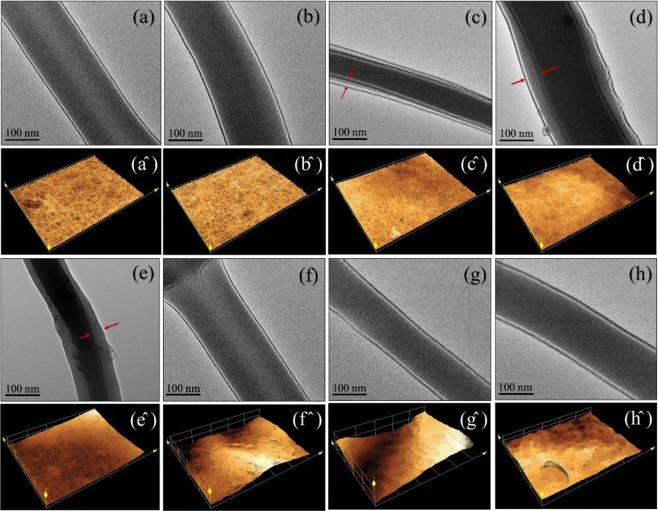
Figure 3.
The TEM images and laser scanning confocal microscopy of uncross-linked PVA-DSs: (a) and (a^), cross-linked PVA-DSs: (b) and (b^), PVA-DSs/PAN-GENs was coated with 0.5% w/w PAN-GENs (c) and (c^), PVA-DSs/PAN-GENs was coated with 1% w/w PAN-GENs (d) and (d^), PVA-DSs/PAN-GENs was coated with 2% w/w PAN-GENs (e) and (e^), PVA-DSs/PAN-GENs was coated with 3% w/w PAN-GENs (f) and (f^), PVA-DSs/PAN-GENs was coated with the 4% w/w PAN-GENs (g) and (g^), PVA-DSs/PAN-GENs was coated with 5% w/w PAN-GENs (h) and (h^).

