Fig. 1.
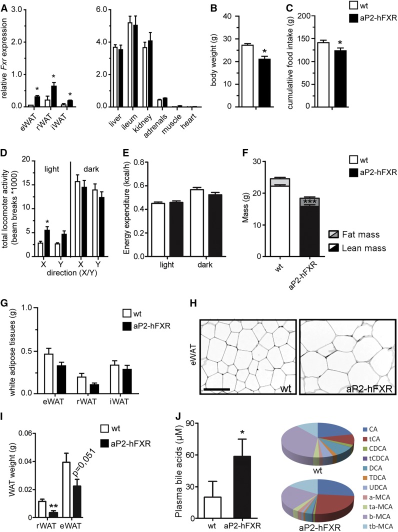
Adipose hFXR-overexpressing mice are small and have hypertrophic adipocytes. A: Quantitative real-time PCR of total FXR expression in WAT depots (epididymal, retroperitoneal, and inguinal), liver, ileum, kidney, adrenals, muscle, and heart of wild-type (wt) and adipocyte-specific FXR overexpressing mice (aP2-hFXR). Body weight (B), cumulative food intake from 6 to 13 weeks of age (C), and locomotor activity (D) of wild-type and aP2-hFXR mice. E: Energy expenditure at 21°C measured over 12 h light/12 h dark phase. F: Lean and fat mass after 13 weeks. WAT depot weights (G) and representative image of eWAT morphology showing adipocyte hypertrophy (H). I: Depot size of rWAT and eWAT 3 weeks after birth of wild-type and aP2-hFXR mice. J: Plasma bile acid composition and total amounts in wild-type and aP2-hFXR mice. Scale bar represents 100 μm. All panels: n = 6–8 per group; data are represented as mean ± SEM; *P < 0.05, **P < 0.01, ***P < 0.001.

