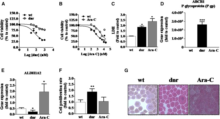
Fig. 1.
Characteristics of chemotherapy-naive HL-60 cells and HL-60 cells grown under dnr and Ara-C selection pressure. A: dnr sensitivity in wt and HL-60/dnr cells. B: Ara-C sensitivity in wt and HL-60/Ara-C cells. Cells (50,000) were seeded in black-walled 96-well plates and exposed to either dnr or Ara-C at the concentrations shown for 72 h. Viability was determined using PI staining. C: LDH levels in HL-60 (wt) and dnr- and Ara-C-resistant counterparts. The assay was conducted in dnr- and Ara-C-free media. D: ABCB1 expression. E: ALDH1A2 expression in wt and dnr- and Ara-C-resistant cells. Gene expression was quantitated by RT-PCR as detailed in Materials and Methods. F: Cellular proliferation rates. Cells (400,000) were seeded in 12-well plates and cultured for 72 h, after which the cell number was determined using automated enumeration and disposable hemocytometers. Dnr and Ara-C were present during the proliferation experiment. G: Cell morphology. Cytospin preparations were stained as detailed in Materials and Methods (200× magnification). HL-60 wt (control, chemotherapy-naive), HL-60/dnr (resistant to 400 nM dnr), and HL-60/Ara-C (resistant to 500 nM Ara-C) cells were used in all experiments.

