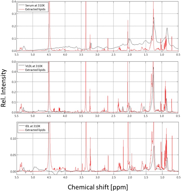
Fig. 3.
1H NMR spectra of intact human serum, VLDL, IDL, and of the corresponding extracted lipids. The 600 MHz 1H NMR spectra of human blood serum (top), VLDL (middle), and IDL (bottom) together with the lipids extracted from the same samples. Samples were contained in synthetic extracellular buffer and measured at 310 K; extracted lipids were dissolved in CDCl3/methanol-d4 (2:1) and measured at 293 K. The spectra were referred to DSS or TMS, respectively, as internal standards. Rel., relative.

