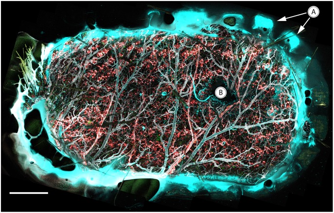
Figure 8.
In vivo confocal tile scan of the complete vacuum window opening attached to immunostained mouse skin. (A) Teeth of the window border lining the tissue, (B) MIP-2 gel positioned between the coverslip and underlying tissue. Color code: Ly6G–cyan, CX3CR1–green, CD31–gray, NG2–red. Scale bar is 400 μm.

