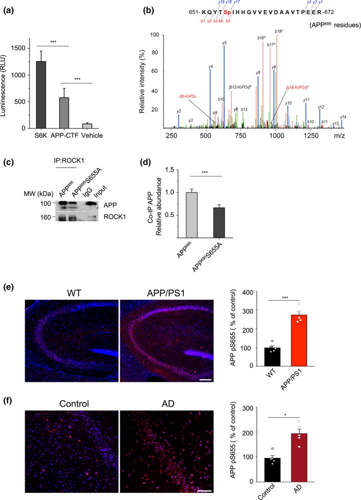
Figure 2.

ROCK1 phosphorylates APP695 at Ser655. (a) ROCK1 activity assay when recombinant ROCK1 protein was incubated with S6K, APP‐CTF, and vehicle. One‐way ANOVA followed by Student–Newman–Keuls test, F 3,28 = 6.28. (b) Representative S655 phospho‐peptide spectrum of APP695. APP‐CTF was incubated with recombinant ROCK1 and examined by LC‐MS/MS after 3 hr. (c) Co‐IP experiments in HEK 293T cells confirmed decreased interaction of ROCK1 with APP after S655A mutation in APP695 plasmid. (d) Quantification of interaction between ROCK1 and APP after APP695 and S655A plasmid transfection in HEK 293T cells. t 4 = 6.67, two‐tailed Student's t test. (e) Immunofluorescence and quantification of phosphorylated APP at S655 (APP pS655) in the hippocampus of APP/PS1 and WT mice (n = 6, t 10 = 8.72). Scale bar, 50 μm. (f) Immunofluorescence and quantification of phosphorylated APP at S655 (APP pS655) in the hippocampus of AD patients and normal control (n = 6, t 10 = 5.8). Scale bar, 50 μm. Data were presented as Mean ± SEM. ***p < 0.001; *p < 0.05
