
Figure 4.
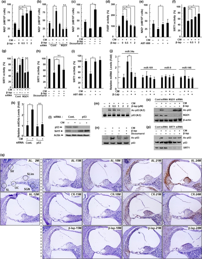
β‐Lap restores the NAD+ and SIRT1 activity in HEI‐OC1 cells. HEI‐OC1 auditory cells were treated with CM in the presence of indicated β‐lap dose for 24 hr (a, d, f, j). Then, we measured the changes in NAD+ levels using a fluorescent NAD+ detection kit (a), PARP activity using the PARP assay kit (d), SIRT1 activity using the SIRT1 assay kit (f), levels of miR‐34a, miR‐181, miR‐9, and miR‐146 by qRT–PCR (j). HEI‐OC1 auditory cells were pretreated with dicoumarol (NQO1 enzymatic inhibitor, 10 μM) (c, h) for 1 hr. These cells were treated with CM with or without β‐lap (1 μM) for 24 hr. Then, NAD+ levels (c) and SIRT1 activity (h) were measured. HEI‐OC1 auditory cells were pretreated with ABT‐888 (PARP inhibitor, 1 μM) (e, i) for 1 hr. These cells were treated with CM for 24 hr. Then, NAD+ levels (e) and SIRT1 activity (i) were measured. HEI‐OC1 auditory cells were transfected with siRNAs against NQO1(b, g) or p53 (k) for 36 hr, and then stimulated with CM for 24 hr in the absence or presence of β‐lap (1 μM). Then, NAD+ level (b), SIRT1 activity (g), and miRNA34a level (k) were measured. Each value represents the mean ± SD (n = 5). *p < .05. HEI‐OC1 auditory cells were transfected with scrambled control siRNA or siRNA against p53 for 36 hr, and then stimulated with CM for 24 hr. Then, Western blot analyses were performed using antibodies for p53, SIRT1, and β‐actin (l). HEI‐OC1 auditory cells were treated with CM in the presence of indicated β‐lap dose for 24 hr. Then, nuclear fractions were prepared and Western blot analyses were performed using antibodies for p53 and ac‐p53 (m). HEI‐OC1 auditory cells were pretreated with dicoumarol for 1 hr. These cells were treated with CM in the presence of indicated β‐lap dose for 24 hr. Then, Western blot analyses were performed using antibodies for p53 and ac‐p53 (n). HEI‐OC1 auditory cells were transfected with scrambled control siRNA or siRNA against NQO1 for 36 hr, and further stimulated with CM for 24 hr in the absence or presence of β‐lap. Then, Western blot analyses were performed using antibodies for ac‐p53, NQO1, and β‐actin (o). HEI‐OC1 auditory cells were transfected with scrambled control siRNA or siRNA against SIRT1 for 36 hr, and further stimulated with CM for 24 hr in the absence or presence of β‐lap. Then, Western blot analyses were performed using antibodies for ac‐p53, p53, and SIRT1 (p). Cochleae were removed from C57BL/6J mice that were fed with either AL, CR diet, or β‐lap‐supplemented diet. Thereafter, the cochleae were decalcified and embedded in paraffin. Next, 5‐μm‐thick sections were prepared. Then, immunohistochemical staining was performed for acetylated p53 in mouse cochleae. All procedures are described in “EXPERIMENTAL PROCEDURES” section. OC, organ of Corti; SG, spiral ganglion neuron; SL, spiral ligament; SV, stria vascularis