
Figure 2.
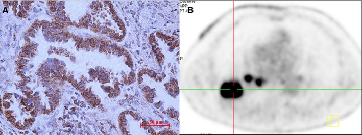
PET imaging of a case with high IDH3a expression. (A) The right inferior lung lobe of a 68‐year‐old man showed an adenocarcinoma with high IDH3a expression (immunohistochemical staining ×400; scale bar, 50 μm). (B) [18F]‐FDG PET/CT scan showed significant accumulation of [18F]‐FDG in the tumor (SUVmax = 15.9, MTV = 10.2 cm3 and TLG = 86.3 cm3). IDH, isocitrate dehydrogenase; SUVmax, maximum standard uptake value; MTV, metabolic tumor volume; TLG, total lesion glycolysis