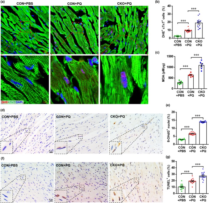
Figure 5.

Cardiac‐specific knockout of Forkhead box O3 (FoxO3) exacerbates paraquat (PQ)‐induced oxidative damage in heart. (a) Representative images of dihydroethidium (DHE) staining showing that PQ‐induced reactive oxygen species (ROS) generation in cardiomyocytes are further enhanced by FoxO3 deletion. (b) Quantification of DHE+ cTnT+ cells in hearts for each condition. Values are presented as mean ± SEM (n = 15 sections from five hearts for each group), ***p < 0.001. (c) Quantification of malondialdehyde concentration in hearts from each group. Values are presented as mean ± SEM (n = 6 hearts for each group), ***p < 0.01. (d) Representative images of 8‐OHdG staining in hearts showing enhanced DNA damage in FoxO3 CKO mice compared with CON mice under PQ‐exposed conditions. (e) Quantification of 8‐OHdG+ cells in hearts for each condition. Values are presented as mean ± SEM (n = 15 sections from five hearts for each group), ***p < 0.001. (f) Representative images of TUNEL staining in hearts showing enhanced apoptosis in FoxO3 CKO mice compared with CON mice under PQ‐exposed conditions. (g) Quantification of TUNEL+ cells in hearts for each condition. Values are presented as mean ± SEM (n = 15 sections from five hearts for each group), ***p < 0.001
