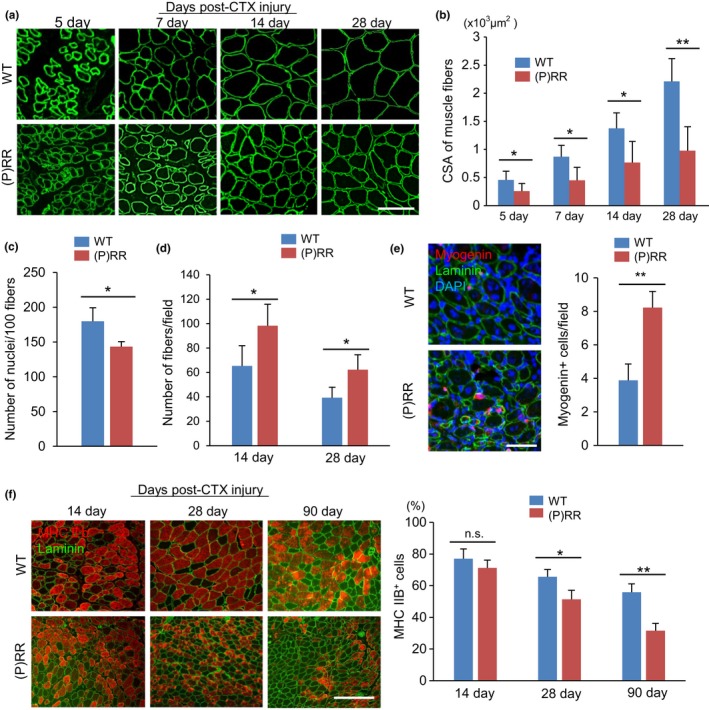
Figure 3.

Histological recovery of skeletal muscle after cardiotoxin (CTX) injection is impaired in (P)RR‐Tg mice. (a) Immunostaining for laminin (green) in cross sections of TA in WT and (P)RR‐Tg mice on day 5, 7, 14, and 28 after CTX injection. Scale bars, 100 mm. (b) Cross‐sectional area of regenerated muscle fibers in WT and (P)RR‐Tg mice on days 5, 7, 14, and 28 after CTX injury (n = 6). (c) Number of nuclei per myofiber in WT and (P)RR‐Tg mice on day 14 after CTX injection (n = 6). (d) Number of central nuclear myocytes per cross section in WT and (P)RR‐Tg mice on days 14 and 28 after CTX injection (n = 6). (e) Immunofluorescent staining for myogenin (red) and laminin (green) in cross sections of TA in WT and (P)RR‐Tg mice on day 7 after CTX injection. Nuclei were labeled with DAPI (blue) (left). Numbers of myogenin‐positive regenerating myocytes in WT and (P)RR‐Tg mice on day 7 after CTX injection (right) (n = 6). Scale bars, 100 mm. (f) Immunofluorescence staining for MHC IIB (red) and laminin (green) in cross sections of TA in WT and (P)RR‐Tg mice on days 14, 28, and 90 after CTX injection (left). Scale bars, 300 mm. Percentage of MHC IIB‐positive myocytes in cross sections of TA after CTX injection (right) (n = 6). Data represent the mean ± SEM. *p < 0.05 and **p < 0.01; n.s., not significant, as determined by the Mann–Whitney U test
