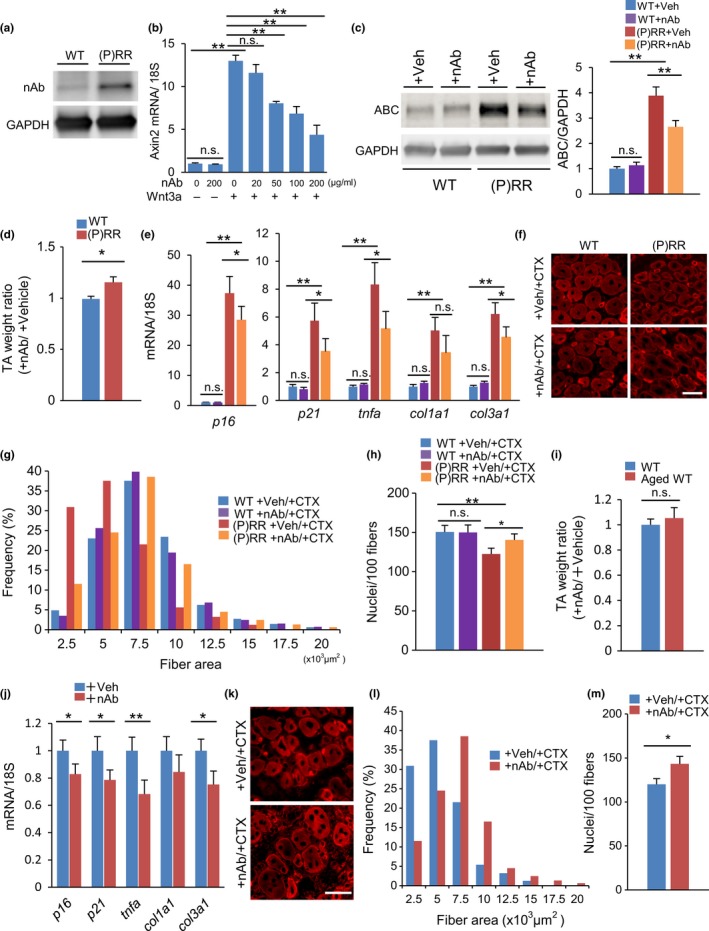
Figure 5.

The anti‐(pro)renin receptor((P)RR) neutralizing antibody (nAb) restores muscle atrophy in (P)RR‐Tg mice and aged mice. (a) Validation of anti‐(P)RR nAb by Western blotting. Protein extract from (P)RR‐Tg mice was used as positive control. (b) Relative expression of axin2 mRNA in Wnt3a‐stimulated C2C12 myoblasts treated with nAb. Expression levels were normalized to those of 18S rRNA and to those in myoblasts subjected to neither Wnt3a nor nAb treatment (n = 7). (c) Western blotting of active β‐catenin (ABC) expression in TA in WT and (P)RR‐Tg mice treated with nAb or vehicle (left) and quantification by densitometry (right) (n = 6). (d) Comparison of the weight ratio of nAb‐treated to vehicle‐treated TA muscle between WT and (P)RR‐Tg mice (n = 7). (e) Relative expression levels of p16, p21, tnfa, col1a1, and col3a1 mRNA in muscles from WT and (P)RR‐Tg mice treated with nAb or vehicle (n = 7). (f) Immunofluorescence staining for embryonic MHC (eMHC) (red) in a cross section of muscle in WT and (P)RR‐Tg mice on day 5 after cardiotoxin (CTX) injection. Scale bars, 50 mm. (g) Cross‐sectional area (CSA) of eMHC‐positive myofibers (n = 7). (h) Number of nuclei per 100 myofibers (n = 7). (i) Comparison of the weight ratio of nAb‐treated to vehicle‐treated TA between young and aged mice (n = 7). (j) Relative expression levels of p16, p21, tnfa, col1a1, and col3a1 mRNA in muscles from aged mice treated with nAb or vehicle (n = 6). (k) Immunofluorescent staining for eMHC (red) in a cross section of muscle in aged mice treated with nAb or vehicle on day 5 after CTX injection. Scale bars, 50 mm. (l) CSA of eMHC‐positive myofibers (n = 7). (m) Number of nuclei per 100 myofibers (n = 7). Data represent the mean ± SEM. *p < 0.05 and **p < 0.01; n.s., not significant, as determined by the Mann–Whitney U test (d,i,j,m) or ANOVA followed by the Bonferroni post hoc correction (b,c,e,h)
