Figure 2.
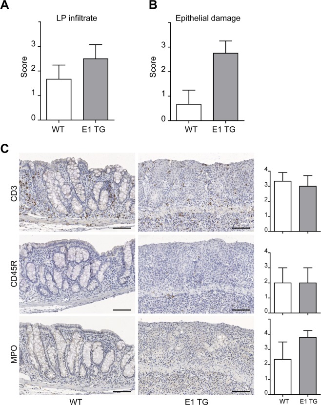
Evaluation of DSS-treated colon with standard IHC. (A) The LP infiltrate status was scored as follow: 0 = absence of infiltrating cells; 1 = poor presence of infiltrating cells; 2 = moderate presence of infiltrating cells; 3 = severe presence of infiltrating cells. (B) Epithelial damage was determined evaluating the integrity of the epithelial layer of the enteric mucosa. This parameter was scored as 0 = absence, 1 = low, 2 = moderate; 3 = severe. (C) Representative images of standard IHC of WT and E1 TG treated colon samples for T (CD3) and B (CD45R/B220) lymphocytes and myeloid cells (MPO). Inflammatory cells were scored counting the number of positive cells within the mucosa (excluding cells of lymphoid follicles): 0 = cell absence; 1 = 1–5 cells; 2 = 6–25 cells; 3 = 26–125 cells; 4 > 125 cells. Bar graphs show mean score values ± s.d. from at least three mice individually tested per group. Scale bar = 100 μm.

