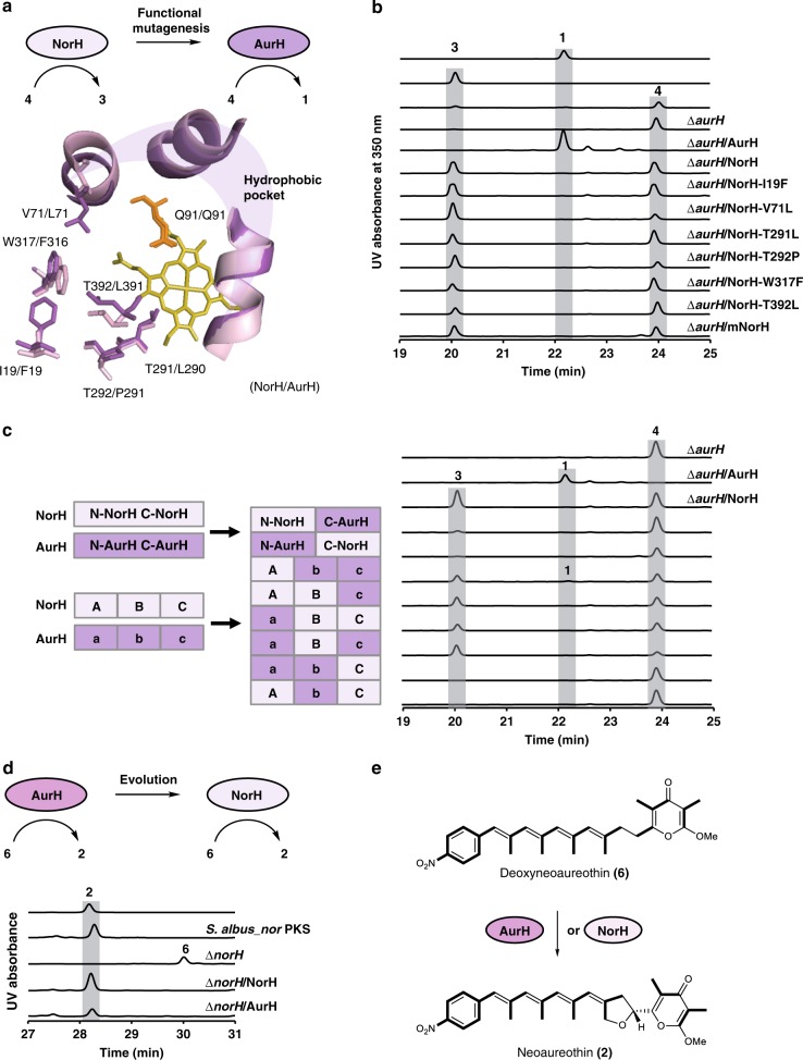
Fig. 4.
Mutagenesis and comparison of oxygenase functions. a Site-directed mutagenesis of NorH. The active center of NorH/AurH model shows a hydrophobic pocket. Residues that may contribute to the different sizes of the cavity are presented in stick. The catalytic residue glutamine (orange) and the cofactor heme (yellow) are indicated. b The HPLC profile of authentic standard of aureothin (1), 7-OH-deoxyaureothin (3), deoxyaureothin (4), and NorH variants. UV detection is at 350 nm. c Schematic presentation of strategies to prepare hybrid NorH/AurH variants, and profiling of biotransformations. The hybrid NorH/AurH variants (left panel) correspond to HPLC profiles of extracts (right panel) of those variants. UV detection at 350 nm. d The HPLC profile of authentic standard of 2 and S. albus mutant strains. e AurH and NorH catalyze the biotransformation from 6 to 2

