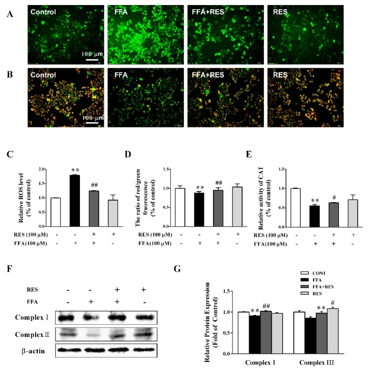
Figure 3.
RES attenuates FFA-triggered oxidative stress and mitochondrial dysfunction in hepatic HepG2 cells. HepG2 cells were pretreated with RES (100 µM) for 6 h and with FFA (100 µM) for 24 h. Then, the cells were stained with 2′,7′-dichlorodihydrofluorescein diacetate (DCFH-DA) to detect intracellular reactive oxygen species (ROS) levels, (A) and (C). (B) and (D) mitochondrial membrane potential (MMP) was examined by JC-1 staining (×200). (E) Intracellular catalase (CAT) activity. (F) Expression levels of mitochondrial complexes, complex I and complex III, in total cell lysates using western blot analysis. β-actin was used as a loading control. (G) Densitometric analysis of the blots shown in (F). Data were presented as the mean ± SD, n ≥ 3. (∗) p < 0.05 and (∗∗) p < 0.01, versus control group; (#) p < 0.05 and (##) p < 0.01, versus FFA group.

