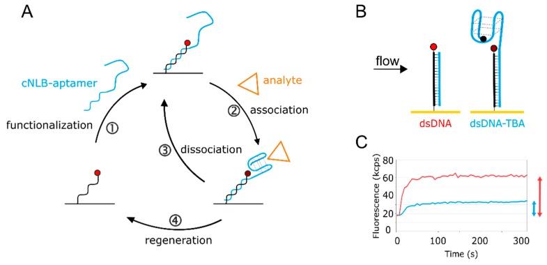
Figure 1.
Aptasensor setup. (A) The aptasensor workflow consisting of ① sensor functionalization, ② + ③ analyte kinetics, and ④ sensor regeneration. The cycle can be automated to continue to bring various targets of interest onto the surface. (B) Scheme of the aptasensor surface. On the left, the immobilized single-stranded nanolever (NL) is functionalized with a complementary sequence (cNL), forming dsDNA. On the right, cNL is extended with the thrombin-binding aptamer (TBA) sequence, carrying a quencher on the distal end. The quencher is optional and enhances the signal. Without quencher, the 5′ guanine of TBA affects the fluorophore, but to a lower extent. (C) Real-time surface functionalization with dsDNA (red) and dsDNA-TBA with quencher (blue). Fluorescence amplitudes reached are marked with respective arrows.

