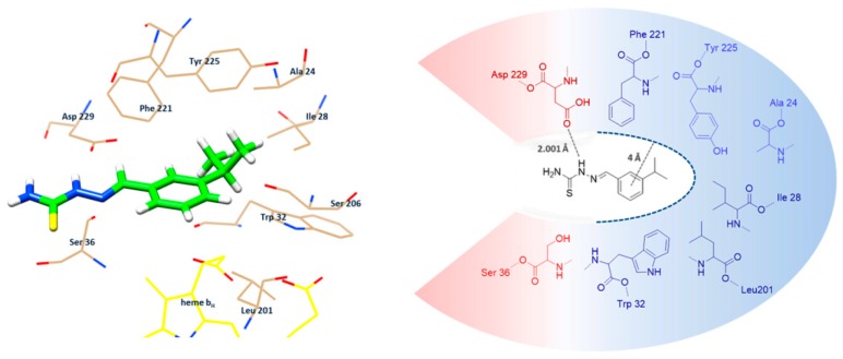
Figure 6.
Representation of the binding mode of mHtcum in the cytochrome bc1 AMY binding site as obtained with the Gold v5.5 software. On the left: best docked pose of mHtcum into the 3BCC binding pocket. On the right: schematic positioning of mHtcum into the 3BCC binding pocket (in red: the hydrophilic area; in blue, the hydrophobic surface).

