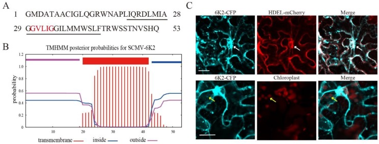
Figure 1.
Subcellular localization of Sugarcane mosaic virus (SCMV)-6K2. (A) The schematic diagram of the amino acids of the SCMV-6K2 protein. GXXXG motif (‘X’ being any amino acid) was highlighted by the red color. The predicted transmembrane domain (TMD) was marked by an underline. (B) Prediction of SCMV-6K2 TMD by TMHMM. The horizontal axis indicates the amino acid position. (C) Subcellular localization of 6K2-CFP in the leaves epidermal cells of N. benthamiana by 48-h post agroinfiltration. White arrows point to endoplasmic reticulum (ER), yellow arrows point to chloroplast. Scale bars, 25 μm.

