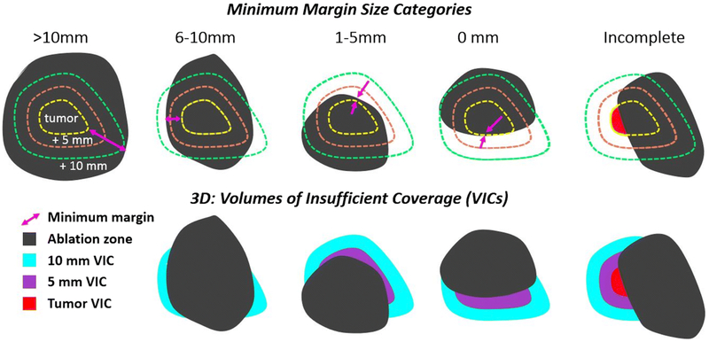
Figure 1.
Various scenarios of the ablation zone’s coverage of the tumor and the tumor plus 5 and 10 mm theoretical margins were divided into five categories (top row), based on the size of the minimum ablation margin (magenta arrows). The bottom row illustrates the three-dimensional assessment metrics, volumes of insufficient coverage (VICs): tumor VIC (red), 5 mm VIC (purple), 10 mm VIC (cyan).

