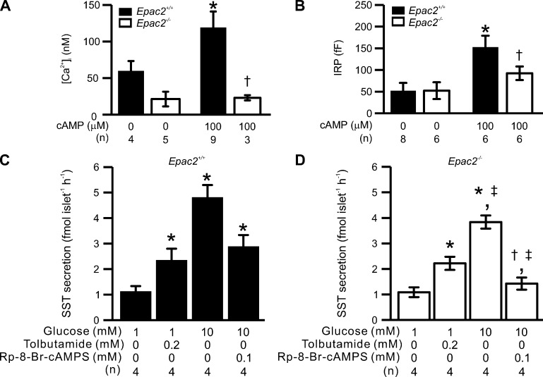
Figure 11.
Ablation of Epac2 (Rapgef4) reduces depolarization-triggered [Ca2+]i, exocytosis and GISS in δ-cells. Somatostatin secretion, depolarization-triggered [Ca2+]i increases, and exocytosis in Epac2-deficient δ-cells. (A) Summary of basal [Ca2+]i measured in the presence and absence of 100 µM cAMP in δ-cells from control (Epac2+/+, filled bars) and Epac2 KO (Epac2−/−, open bars) mice. *, P < 0.05 vs. basal [Ca2+]i measured in δ-cells without cAMP of the same genotype; †, P < 0.05 vs. basal [Ca2+]i measured in Epac2+/+ δ-cells infused with 100 µM cAMP. (B) Summary of IRP determined in Epac2+/+ (filled bars) and Epac2−/− (open bars) δ-cells with treatments as indicated below. *, P < 0.05 vs. IRP measured in δ-cells without cAMP of the same genotype; †, P < 0.05 vs. IRP measured in Epac2+/+ δ-cells infused with 100 µM cAMP. (C and D) Somatostatin secretion measured in islets from Epac2+/+ (C, filled bars) and Epac2−/− mice (D, open bars) under the indicated experimental conditions. *, P < 0.05 vs. 1 mM glucose alone of the same genotype; †, P < 0.05 vs. tolbutamide alone of the same genotype; ‡, P < 0.05 vs. responses under the same condition in Epac2+/+ islets. In A–D, data are presented as mean values ± SEM of indicated numbers of independent experiments (n) from two to six animals for each experimental condition. In A and B, experiments were conducted using the standard whole-cell technique.

