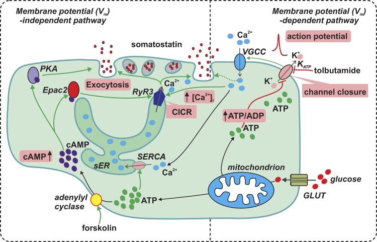
Figure 15.
Glucose stimulates somatostatin secretion through both membrane potential-dependent and -independent pathways. Membrane potential (Vm)–dependent pathway: glucose (large red particles) metabolism increases δ-cell intracellular ATP (green particles) at the expense of ADP and consequently inhibits the KATP channels (sensitive to tolbutamide). This leads to membrane depolarization and increase in electrical activity that opens voltage-gated Ca2+ channels (VGCC) to allow influx of extracellular Ca2+ (blue particles) to increase [Ca2+]i. This exerts a relatively weak stimulation on δ-cell exocytosis (dotted line). Vm-independent pathway: high glucose stimulates somatostatin secretion by promoting CICR in δ-cells. First, the glucose-induced stimulation of ATP production increases ER luminal Ca2+ content through SERCA pump. Second, glucose metabolism elevates [cAMP]i (dark blue particles), a second messenger which activates PKA and Epac2. Whereas PKA directly potentiates δ-cell exocytosis, the effect of Epac2 on exocytosis is mediated through the activation of RYR3 and sensitization of CICR. The cAMP-dependent effects are mimicked by the adenylyl cyclase activator forskolin. The Vm-dependent and -independent pathways operate synergistically leading to a full stimulatory effect of somatostatin secretion by glucose. The key events (increase in ATP/ADP ratio, KATP channel closure, action potential firing, increase in [cAMP]i, CICR, increase in [Ca2+]i, and exocytosis) are highlighted in pink boxes. Green arrows indicate stimulation, and the red blind-ended arrows indicate inhibition. sER, smooth endoplasmic reticulum; GLUT, glucose transporter.

