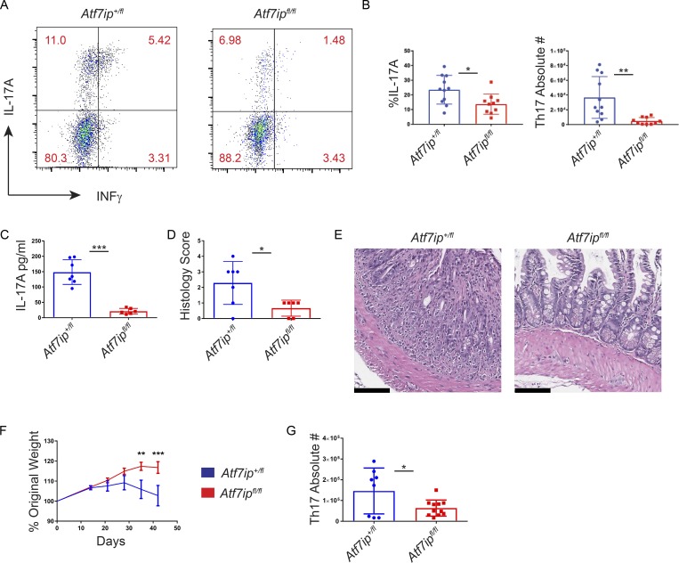
Figure 2.
Atf7ip deletion attenuates colitis in vivo. (A–E) CD4-Cre/Atf7ip+/fl (Atf7ip+/fl) and CD4-Cre/Atf7ipfl/fl (Atf7ipfl/fl) mice were injected intraperitoneal with anti-CD3 antibodies and analyzed 48 h after injection. (A) Flow cytometric analysis of CD4+IL17A+INFγ+ small intestine IELs. (B) Percentage and absolute numbers intraepithelial CD4+ T cells in the small intestine expressing IL-17A. (C) Serum IL-17A levels measured by ELISA. (D) Histological score of the small intestine. (E) H&E staining of the small intestine. Bars, 100 µm. (F) Weight change in Rag1−/− recipients of RBhi naive T cells from either CD4-Cre/Atf7ip+/fl or CD4-Cre/Atf7ipfl/fl mice measured on day 0, 14, 21, 28, 35, and 42. Results are the combination of two experiments with 13 mice per genotype. (G) Absolute number of CD4+IL17A+ T cells in the colonic lamina propria of Rag1−/− mice. Each data point in B–D and G represents an individual mouse. Data are the combination of three (B) or two (C, D, F, and G) independent experiments with three to four mice per group in each experiment. Error bars in B–D and G are mean with SD. Error bars in F are SEM. *, P < 0.05; **, P < 0.01; ***, P < 0.001; significance by Student’s t test (B, C, and G); Mann–Whitney nonparametric test (D); and two-way ANOVA followed by multiple t tests using the Holm–Sidak method (F).

