Figure 3.
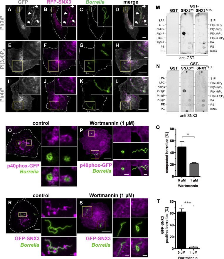
SNX3 binds to PI(3)P and colocalizes with PI(3)P at B. burgdorferi–containing phagosomes. (A–L) Confocal micrographs of macrophages coexpressing fluorescent probes for PI(3)P (A), PI(3,4)P2 (E), or PI(4)P (I), and coexpressing RFP-SNX3 (B, F, and J), with internalized borreliae stained by antibodies (C, G, and K). Yellow boxes indicate areas shown enlarged as insets. Arrows indicate enrichments of PI(3)P and RFP-SNX3. Scale bars: 10 µm, and 1 µm for insets. (M and N) Binding of GST-fused SNX3 WT and SNX3-Y71A mutant on phosphatidylinositol-spotted strips incubated with GST alone or GST-SNX3 WT or GST-SNX3-Y71A mutant and developed with anti-GST antibody (M) or anti-SNX3 antibody (N). LPA, lysophosphatidic acid; LPC, lysophosphocholine; PA, phosphatidic acid; PC, phosphatidylcholine; PE, phosphatidylethanolamine; PS, phosphatidylserine; S1P, sphingosine 1-phosphate. (O–T) PI3K activity regulates compaction of borreliae. (O, P, R, and S) Confocal micrographs of macrophages expressing PI(3)P sensor p40phox-GFP (O and P) or GFP-SNX3 (R and S), with internalized borreliae stained by antibodies, with merges. Cells were treated with DMSO (0.1%) as control (O and R) or with PI3K inhibitor Wortmannin (1 µM; P and S); yellow boxes indicate areas shown enlarged as insets. Scale bars: 10 µm, and 1 µm for insets. (Q and T) Statistical evaluation of borreliae compaction in cells expressing p40phox-GFP (Q) or of GFP-SNX3 localization to borreliae phagosomes (T); cells were treated with Wortmannin or DMSO as control. n = 3 (3 × 30 cells). For specific values, see Table S1. Two-tailed Student’s t test; *, P < 0.05; ***, P < 0.001. Error bars: mean ± SEM.

