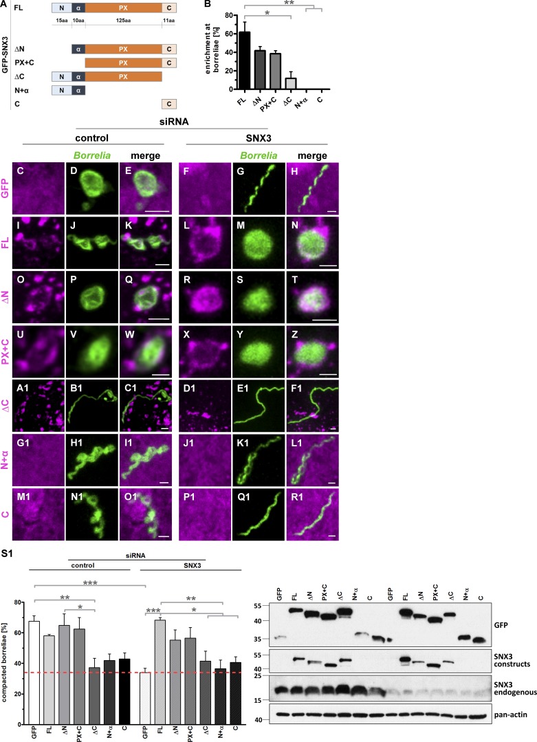
Figure 5.
The C-terminal region of SNX3 is crucial for localization to phagosomes and borreliae compaction. (A) Graphic representation of SNX3 regions used in respective GFP-fused constructs: the N-terminal region (N), followed by an α-helical region (α), a PX domain, and a C-terminal region (C). (B) Enrichment of SNX3 constructs at borreliae phagosomes, with total number of phagosomes set to 100%. Values are presented as mean ± SEM, with 3 × 30 phagosomes analyzed. One-way ANOVA; *, P < 0.05; **, P < 0.01. For specific values, see Table S1. (C–R1) Colocalization of GFP-fused SNX3 constructs at borreliae phagosomes. Confocal micrographs of macrophages transfected with respective constructs (magenta), with borreliae stained by B. burgdorferi–specific antibody (green), and merges. Cells were treated with control siRNA or SNX3-specific siRNA, as indicated. Scale bars: 1 µm. (S1) Evaluation of borreliae phagosome compaction in macrophages expressing siRNA-insensitive SNX3 constructs. Top: Statistical evaluation of borreliae compaction in cells treated with control siRNA or SNX3-specific siRNA and expressing indicated constructs. n = 4 (4 × 30 cells). Dotted red line indicates level of borreliae compaction in SNX3-depleted control (GFP-expressing) cells. For specific values, see Table S1. One-way ANOVA; *, P < 0.05; **, P < 0.01; ***, P < 0.001. Bottom: Western blots of respective macrophage lysates, developed with GFP-specific or SNX3-specific antibody, the latter used for detection of both GFP-fused and endogenous forms of SNX3, with pan-actin as loading control. Molecular weight is indicated in kilodaltons on the left.

