Figure 5.
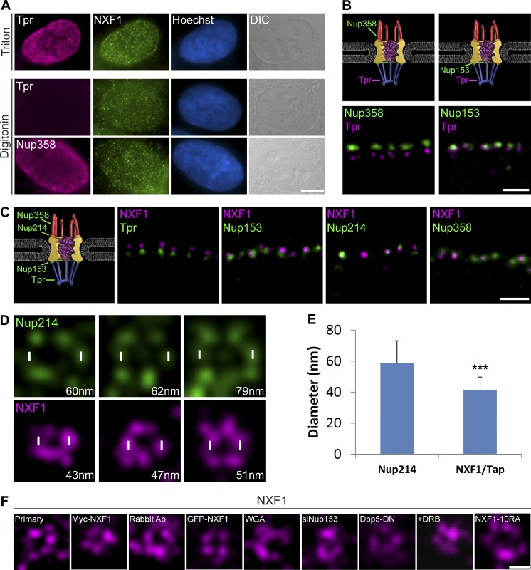
NXF1 is situated mainly on the cytoplasmic side of the NPC. (A) U2OS cells were permeabilized with either Triton X-100 (top) or digitonin (bottom) and coimmunostained with anti-NXF1 together with anti-Tpr (nuclear basket marker) or anti-Nup358 (cytoplasmic side marker). Hoechst DNA stain, blue; differential interference contrast (DIC), gray. Scale bar, 5 µm. (B) STED images of regions of the nuclear envelope of U2OS cells immunostained with antibodies to Tpr and Nup358 (left) or Nup153 (right). (C) Representative STED images of regions of the nuclear envelope immunostained with antibodies to NXF1 together with antibodies against Tpr, Nup153, Nup214, or Nup358 (n = 780, 1,428, 556, and 900 NPCs; 13, 26, 16, and 16 cells, respectively). Measurements were performed in three independent experiments. Scale bars, 0.5 µm. (D) Representative STED images showing a top view of either Nup214 (top) or NXF1 (bottom; in different experiments) within single NPCs imaged at the nuclear surface. Diameter of the circular pattern created by the proteins is shown in the bottom-right corner of each image (script NPC_diameter in the online supplemental material). (E) Plot showing the average diameter of the circular patterns of Nup214 and NXF1. Error bars represent SD. See Materials and methods for the number of NPCs and cells shown. ***, P < 0.001. Measurements were performed in three independent experiments. (F) Representative STED images showing a top view of NXF1 staining in single NPCs imaged at the nuclear surface under the following conditions: using a fluorescently labeled primary anti-NXF1 antibody; Myc-tagged NXF1; rabbit antibody to NXF1; GFP-NXF1; export blocks WGA, siNup153, and Dbp5-DN; DRB treatment; and NXF1-10RA.

