Figure 4.
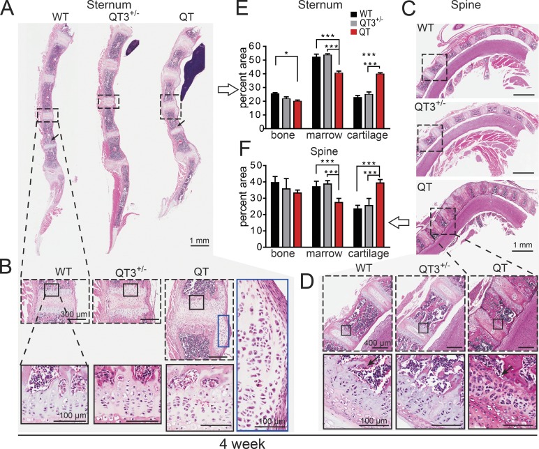
TIMPs are required for normal morphology of axial bone. (A–D) H&E-stained sagittal plane section of sternum (A and B) and spine (C and D; cervical and thoracic vertebrae) from 4-wk old mice. (B and D) Marked areas compare a single cartilaginous joint bordered by two growth plates. Further magnification exhibits cellular organization in growth plate cartilage and laterally migrated cartilage of QT sternum. Black arrow indicates loss of trabecular bone. (E and F) Histomorphometric quantification of percentage of cartilage, bone, and marrow in sternums and spine (7–8 wk-old; n = 3–6/group). ANOVA with Bonferroni’s multiple comparison test assessed significance for each tissue. *, P < 0.05; ***, P < 0.001.

