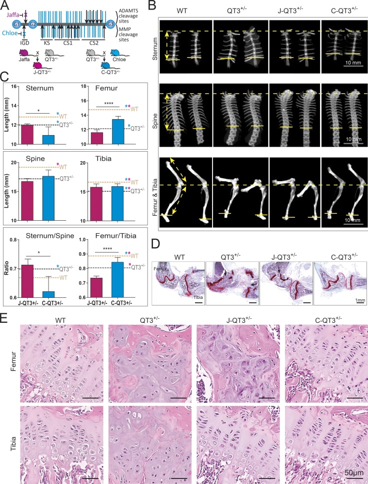
Figure 5.
Metalloprotease-resistant aggrecan rescues long bone proportionality. (A) Structure of aggrecan and the Chloe/Jaffa knock-in mutation. The aggrecan protein core (gray) contains three globular regions (G1–G3) and has ∼100 glycosaminoglycan chains (purple) attached. MMP and ADAMTS cleavage sites are indicated by black arrows. Disruption of specific cleavage sites by knock-in mutations are indicated by an x. Schematic of breeding strategies to generate J-QT3+/− and C-QT3+/− mice. CS1, chondroitin sulfate domain 1; CS2, chondroitin sulfate domain 2; KS, keratan sulfate. (B) Representative micro-CT images used to measure the length of sternum, the spine, and the leg bones of 8-wk old WT, QT3+/−, C-QT3+/−, and J-QT3+/−. Sternum (upper panel), spine (middle panel), and leg bones (lower panel). Arrows indicate measurements as described in legend of Fig. 1 H. (C) Quantification of axial and appendicular bones length in 8-wk-old WT, QT3+/−, C-QT3+/−, and J-QT3+/−. The sternum, spine, femur, and tibia are all shorter in the QT3+/− mouse compared with WT controls (dashed lines: WT, orange; QT3+/−, gray). C-QT3+/− mice have shorter sternums. Sternum:thoracic spine ratio indicates the effect was more pronounced in sternum than spine. C-QT3+/− partially rescued the shortened QT3+/− femur length, but J-QT3+/− mice could not. The femur:tibia ratio reflects that C-QT3+/− mice partially restored the leg bone compared with QT3+/− (n = 5–7/each group). Mean values of each dataset are plotted in graphs with error bars representing SEM. Datasets were compared by unpaired t test, *, P < 0.05; ****, P < 0.0001. Dunnett’s test P < 0.05 when WT or QT3+/− compared with C-QT3+/− (blue*) or J-QT3+/− (pink*). (D) Safranin O–stained knee joints of WT, QT3+/−, C-QT3+/−, and J-QT3+/− mice at 8 wk of age. Black arrows show growth plate closure in femur and tibia of QT3+/− mice; femur growth plate is rescued in the C-QT3+/− but not in J-QT3+/−, while the tibia growth plate is rescued by both mutant aggrecan (4×/0.5-NA objective). (E) H&E-stained femur and tibia growth plates of WT, QT3+/−, C-QT3+/−, and J-QT3+/− mice (8-wk-old) chondrocyte disorganization in the growth plates of QT3+/− femur and tibia, J-QT3+/− femur but not in the C-QT3+/− mice. Scale bar = 50 µm; 20×/0.5-NA objective.

