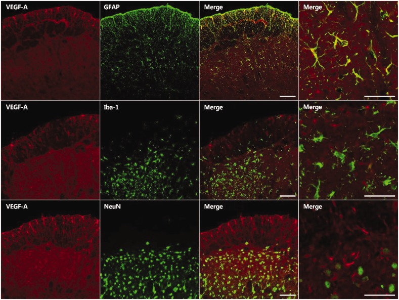
Figure 2.
Double immunofluorescence staining for VEGF-A (red) and either GFAP, an astrocyte marker (green); Iba-1, a microglial marker (green); or NeuN, a neuronal marker (green) on POD 5. The VEGF-A immunoreactive cells primarily colocalized with GFAP. Scale bar, 50 μm.VEGF-A: vascular endothelial growth factor-A.

