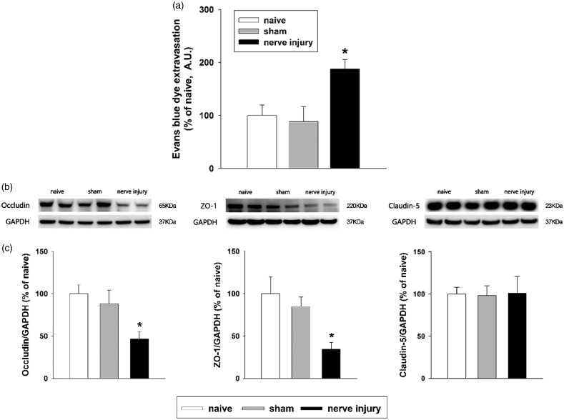
Figure 6.
Evaluation of blood-brain barrier permeability after inferior alveolar nerve injury in rats. (a) High levels of extravasated Evans blue dye were evident on POD 5 in the inferior alveolar nerve injury group compared to the sham-treated group. There were eight animals in each group. *P < 0.05, sham vs. nerve injury group. (b and c) Western blot analysis revealed significantly decreased occludin and ZO-1 expression in the medullary dorsal horn on POD 5. Inferior alveolar nerve injury did not alter the claudin-5 expression level. GAPDH was used as a loading control. There were eight animals in each group. *P < .05, sham vs. nerve injury group. GAPDH: glyceraldehyde 3-phosphate dehydrogenase.

