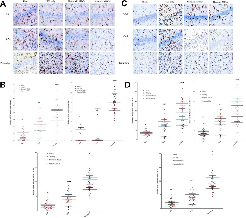
Figure 4.
Photos of histochemical staining and quantity results at different time points after treatment in the hippocampus and penumbral region. (a) and (b) At day 3, more glial fibrillary acidic protein (GFAP)+ve cells were observed in both the hippocampus and penumbral region in the hypoxic mesenchymal stem cell (MSC) group (CA1: *p = 0.036 vs traumatic brain injury (TBI) only, #p = 0.018 vs TBI only, ^p = 0.028 vs sham group; CA3: *p = 0.050 vs TBI only, #p = 0.047 vs TBI only, ^p = 0.049 vs sham group; penumbra: *p = 0.008 vs TBI only, #p = 0.050 vs TBI only, ▪p = 0.050 vs normoxic MSCs, ^p = 0.031 vs sham group). At day 7, the increased number of GFAP-positive cells was only found in penumbra (*p < 0.001 vs TBI only, #p = 0.033 vs TBI only, ▪p = 0.045 vs normoxic MSC, ^p = 0.037 vs sham). More GFAP+ve cells were found in the sham group in hippocampus (CA1: ^p < 0.001; CA3: ^p < 0.001). At day 14, less astrocytes infiltration was found in the hippocampus in hypoxic MSCs group (CA1: *p = 0.038 vs TBI only; CA3: *p < 0.001 vs TBI only, ▪p < 0.001 vs normoxic MSCs, #p = 0.041 vs TBI only). (c)-(d) At day 3, more microglia were activated after TBI in both hippocampus and penumbra (CA1: ^p < 0.001 vs sham group; CA3: ^p < 0.001 vs sham group; penumbra, ^p < 0.001 vs sham group). Fewer ionized calcium binding adapter molecule (Iba)+ve cells were found in the hypoxic MSC group in CA3 and penumbra (CA3: *p = 0.035 vs TBI only; penumbra: *p < 0.001 vs TBI only, ▪p = 0.002 vs normoxic MSCs). Normoxic MSC treatment also reduced the number of Iba+ve cells at CA3 and penumbra (CA3: #p = 0.034; penumbra, #p = 0.036). At day 7, fewer microglia were found in the hypoxic MSC group in both the hippocampus and penumbra (CA1: *p = 0.036 vs TBI only; CA3: *p = 0.040 vs TBI only; penumbra: *p < 0.001 vs TBI only, ▪p = 0.035 vs normoxic MSCs). Normoxic MSC treatment reduced microglia activation at CA1 and penumbra significantly (CA1: #p = 0.011; penumbra: #p = 0.037). Both normoxic and hypoxic MSCs reduced the number of microglia at day 14, no significant difference was found between the two treatments (CA1: *p = 0.043, #p = 0.010; CA3: *p = 0.029, #p = 0.043; penumbra: *p < 0.001, #p < 0.001). (e)-(f) Number of cell death increased significantly after TBI from day 3 to day 14 in both the hippocampus and penumbra (^p < 0.05 vs sham). At day 3, cell death was reduced remarkably in the hypoxic MSC group in both the hippocampus and penumbra (CA1: *p = 0.014 vs TBI only; CA3: *p = 0.005 vs TBI only; penumbra: *p = 0.013 vs TBI only, ▪p = 0.022 vs normoxic MSCs). Normoxic MSCs reduced cell death at the hippocampus only (CA1: #p = 0.024; CA3: #p = 0.038). At day 7, both normoxic

