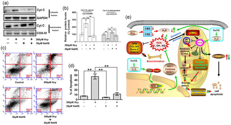
FIGURE 6.
NaHS decrease Hcy-mediated osteoblast apoptosis. (a) Expression of Cyt C in cytosolic fraction and mitochondria fraction which indicated the releasing of Cyt C during cell apoptosis. (b) Quantitation by Image-Pro Plus 6.0 and displayed as relative optical density units after being standardized against housekeeping gene bands, results were normalized with control group. (c) Annexin V/PI staining and flow cytometry to show cell apoptosis. (d) Quantification of flow cytometry to show the ratio of cell apoptosis. (e) Schematic of proposed hypothetical mechanism of NaHS mediated recovery of osteoblast function via mitochondrial biogenesis in Hcy-treated condition. Values are means ± SD, n = 3, *p < 0.05, **p < 0.01 between two compared group. Cyt C: cytochrome c; Hcy: homocysteine; NaHS: sodium hydrogen sulfide; PI: propidium iodide

