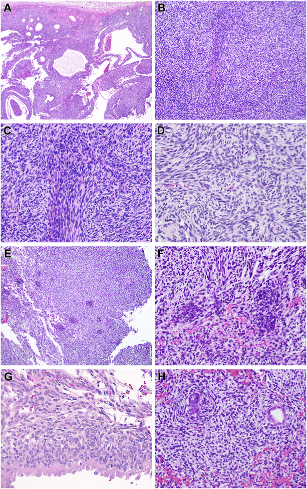
Figure 1. Morphologic features of renal case 1 with MEIS1-NCOA2 fusion (index case).
This neoplasm had a dominant cystic appearance at low power (A), created by a solid spindle cell neoplasm associated with entrapped cystically-dilated renal tubules. The neoplasm had a nodular low power appearance (B) created by centrally located spindle cells with more abundant eosinophilic cytoplasm separated by primitive spindle to epithelioid cells with scant cytoplasm (C). The cytology in these areas overlapped with that of monophasic synovial sarcoma. The centrally located spindle cells demonstrated whorling (D). Small primitive cells arranged in clusters (E) and vague whorling similar to the spindle cells (F). The primitive neoplastic cells were cytologically distinct from the entrapped, cystically-dilated native renal tubular epithelium, which had more open, vesicular chromatin and hobnail appearance (G). In focal areas the spindle cells encircled native tubules in a concentric, “onion-skin” pattern (H).

