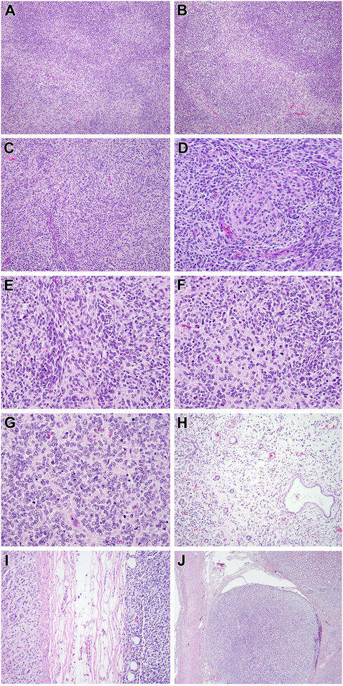
Figure 2. Morphologic features of renal case 2 with MEIS1-NCOA2 fusion.
This solid cellular neoplasm had distinctive nodular variations in cellularity as evident at low power (A). In some areas, the nodularity was created by hypocellular fibrous zones separating more cellular nodules (B). The most consistent pattern, as seen in case 1, was that of centrally located spindle cells with eosinophilic cytoplasm forming whorls, surrounded by smaller cells with less cytoplasm (C, D); the smaller cells were packed in tight clusters, often centered on capillaries (E). Mitotic figures were readily identifiable in both components (F). In more cellular areas, the appearance resembles that of a small blue round cell tumor such as a primitive neuroectodermal tumor (PNET) with Homer Wright rosettes (G). This solid cellular neoplasm focally permeated between native renal tubules at its periphery but a significant cystic component was absent (H). The neoplasm invaded through the renal capsule to approach the adrenal gland (I) and showing invasion into the renal sinus (J).

